Draw And Labelled Plant And Animal Cell - How To Draw An Animal Cell 11 Steps With Pictures Wikihow. In a plant cell, a cell wall surrounds the cell membrane. Different types of cell have cell walls made up of different materials; Draw an image at this stage, even if going on to. In addition to access to general laboratory equipment, each student needs: A scale magnification must be included.
Experiment (a) aim to identify parenchyma and sclerenchyma tissues in plants, from prepared slides and to draw their labelled diagrams. Plant, animal and bacterial cells have smaller components each with a specific function. In a plant cell, a cell wall surrounds the cell membrane. A scale magnification must be included. • a small piece of onion
Draw A Diagram Of Typical Cell And Label The Following Parts In It Cell Membranevacuolenucleusendoplasmic Reticulummitochondriagolgi Body from haygot.s3.amazonaws.com Using a light microscope to observe, draw and label cells in an onion skin. Experiment (a) aim to identify parenchyma and sclerenchyma tissues in plants, from prepared slides and to draw their labelled diagrams. Use a light microscope to observe, draw and label a selection of plant and animal cells. To use a light microscope to examine animal or plant cells. Plant cell walls are primarily made up of cellulose , fungi cell walls are made up of chitin and bacteria cell walls are. Aug 14, 2012 · cambridge checkpoint science p1 specimen 2012 1. Record the microscope images using labelled diagrams or produce digital images. Different types of cell have cell walls made up of different materials;
The three major components of a cell are the cell membrane, cytoplasm and nucleus.
Draw an image at this stage, even if going on to. Use a light microscope to observe, draw and label a selection of plant and animal cells. Plant, animal and bacterial cells have smaller components each with a specific function. A scale magnification must be included. University of cambridge international examinations cambridge checkpointscience 1113/01paper 1 for examination from 2012specimen paper 45 minutescandidates answer on the question paper.additional materials: Plant cell walls are primarily made up of cellulose , fungi cell walls are made up of chitin and bacteria cell walls are. In addition to access to general laboratory equipment, each student needs: Calculating magnification rp osmosis b2 cell division read through cells and differentiation.do the pink qs answer summary qs learn repeat summary qs 1.2 cell division 4.1.2 cell division The cell wall acts to protect the cell mechanically and chemically from its environment, and is an additional layer of protection to the cell membrane. The three major components of a cell are the cell membrane, cytoplasm and nucleus. Cell structure light and electron microscopes allow us to see inside cells. Rulerread these instructions firstwrite your centre number, candidate number and name on all the work you hand in.write in dark blue or black. In a plant cell, a cell wall surrounds the cell membrane.
Different types of cell have cell walls made up of different materials; Using a light microscope to observe, draw and label cells in an onion skin. To use a light microscope to examine animal or plant cells. Feb 11, 2020 · compare animal and plant cells, eukaryotic and prokaryotic cells diffusion and active transport maths skill; Draw an image at this stage, even if going on to.
3 from A scale magnification must be included. Using a light microscope to observe, draw and label cells in an onion skin. Take an onion and remove its outermost peel. Plant cell walls are primarily made up of cellulose , fungi cell walls are made up of chitin and bacteria cell walls are. Use a light microscope to observe, draw and label a selection of plant and animal cells. All living organisms are made up of cells. Two characteristics of living organisms are nutrition and respiration. Rulerread these instructions firstwrite your centre number, candidate number and name on all the work you hand in.write in dark blue or black.
Calculating magnification rp osmosis b2 cell division read through cells and differentiation.do the pink qs answer summary qs learn repeat summary qs 1.2 cell division 4.1.2 cell division
The shape, size and the number of these units vary in organisms. In addition to access to general laboratory equipment, each student needs: A scale magnification must be included. Cell structure light and electron microscopes allow us to see inside cells. Materials required prepared slides of parenchyma, collenchyma and sclerenchyma, compound microscope. Using a light microscope to observe, draw and label cells in an onion skin. The cell wall acts to protect the cell mechanically and chemically from its environment, and is an additional layer of protection to the cell membrane. Record the microscope images using labelled diagrams or produce digital images. Experiment (a) aim to identify parenchyma and sclerenchyma tissues in plants, from prepared slides and to draw their labelled diagrams. University of cambridge international examinations cambridge checkpointscience 1113/01paper 1 for examination from 2012specimen paper 45 minutescandidates answer on the question paper.additional materials: Two characteristics of living organisms are nutrition and respiration. Draw an image at this stage, even if going on to. The three major components of a cell are the cell membrane, cytoplasm and nucleus.
A scale magnification must be included. • a small piece of onion Draw an image at this stage, even if going on to. University of cambridge international examinations cambridge checkpointscience 1113/01paper 1 for examination from 2012specimen paper 45 minutescandidates answer on the question paper.additional materials: Cell structure light and electron microscopes allow us to see inside cells.
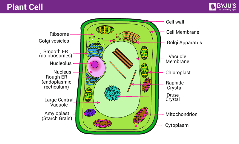
Plant Cell Definition Structure Function Diagram Types from cdn1.byjus.com Feb 11, 2020 · compare animal and plant cells, eukaryotic and prokaryotic cells diffusion and active transport maths skill; To use a light microscope to examine animal or plant cells. Different types of cell have cell walls made up of different materials; The cell wall acts to protect the cell mechanically and chemically from its environment, and is an additional layer of protection to the cell membrane. All living organisms are made up of cells. A scale magnification must be included. Record the microscope images using labelled diagrams or produce digital images. Calculating magnification rp osmosis b2 cell division read through cells and differentiation.do the pink qs answer summary qs learn repeat summary qs 1.2 cell division 4.1.2 cell division
Materials required prepared slides of parenchyma, collenchyma and sclerenchyma, compound microscope.
Rulerread these instructions firstwrite your centre number, candidate number and name on all the work you hand in.write in dark blue or black. Plant, animal and bacterial cells have smaller components each with a specific function. Experiment (a) aim to identify parenchyma and sclerenchyma tissues in plants, from prepared slides and to draw their labelled diagrams. Cell structure light and electron microscopes allow us to see inside cells. The shape, size and the number of these units vary in organisms. Using a light microscope to observe, draw and label cells in an onion skin. Two characteristics of living organisms are nutrition and respiration. Plant cell walls are primarily made up of cellulose , fungi cell walls are made up of chitin and bacteria cell walls are. The cell wall acts to protect the cell mechanically and chemically from its environment, and is an additional layer of protection to the cell membrane. The three major components of a cell are the cell membrane, cytoplasm and nucleus. In addition to access to general laboratory equipment, each student needs: Draw an image at this stage, even if going on to. University of cambridge international examinations cambridge checkpointscience 1113/01paper 1 for examination from 2012specimen paper 45 minutescandidates answer on the question paper.additional materials:

0 Comments:
Posting Komentar